SphynxVision makes its debut at WMIC 2026
SphynxVision was introduced to the molecular imaging community at WMIC 2026 in Salt Lake City, with live demonstrations and four studies presented using the platform.
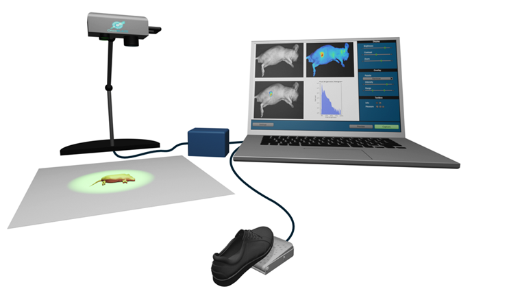
SphynxVision enables real-time, flexible preclinical fluorescence imaging under ambient light — designed for researchers who need to image during interventions, not only after them.
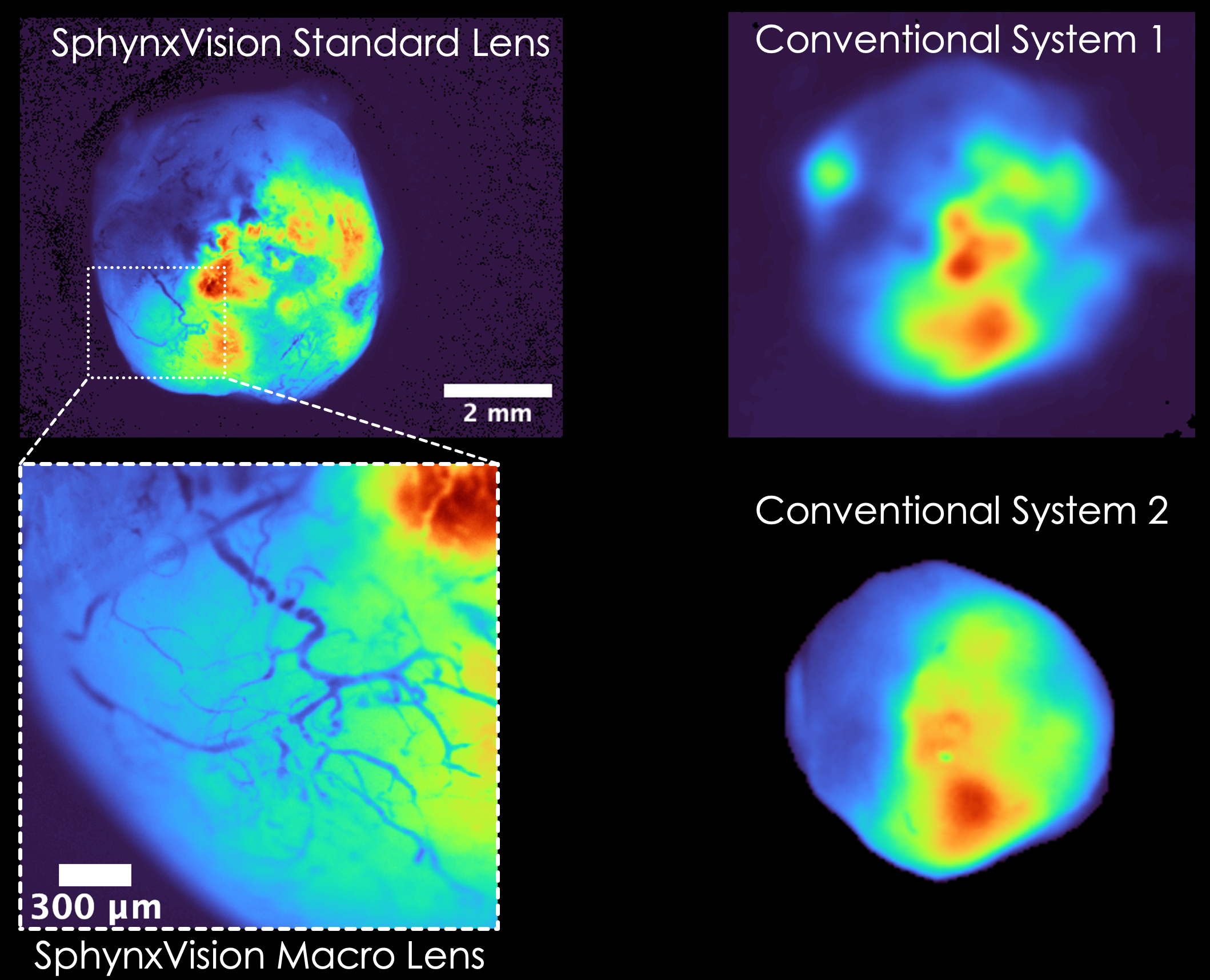
SphynxVision was introduced to the molecular imaging community at WMIC 2026 in Salt Lake City, with live demonstrations and four studies presented using the platform.
OncoBLAZE filed a provisional patent application on innovative fluorescence imaging system named SphynxVision (https://sphynx-vision.com) on February 2026.
A recent LinkedIn post by SphynxVision shows how researchers can use fluorescence for preclinical image-guided surgery during tumor resection.
Existing imaging facilities are powerful, but they often force researchers into scheduled access, closed-chamber workflows, animal transfer, and endpoint-style imaging. SphynxVision gives teams a bench-side, open-access way to image dynamic fluorescence during the study itself.
Proprietary light source and image processing algorithm.
Image when the study is ready instead of waiting for core facility schedules, chamber-based workflows, or shared instrument availability.
Support dosing, heating, perfusion, surgery, and device-assisted workflows without locking the animal inside a closed chamber.
Capture real-time fluorescence kinetics that may be missed by endpoint imaging after transfer to a closed imaging system.
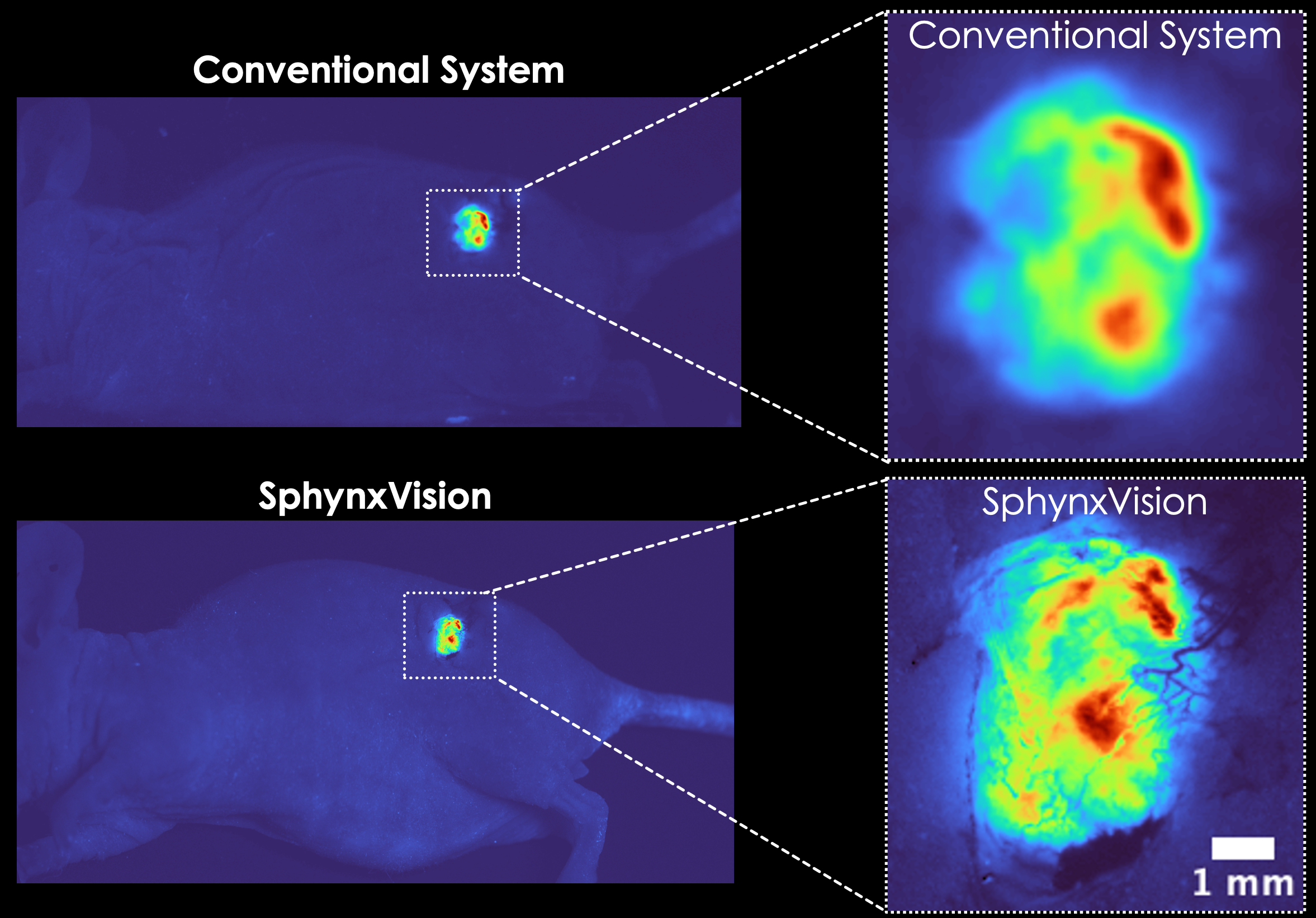
Visualize fluorescence response during the experiment with live display and fast acquisition.
No need for an enclosed chamber for flexible workflows.
Support for visible-to-NIR fluorescence imaging applications.
Fluorescence overlay on the background image in real time.
Capture time series for pharmacokinetics, perfusion, and angiography studies supporting multiple frames per second.
Save imaging settings, wavelength metadata, and calibration values with acquired data.
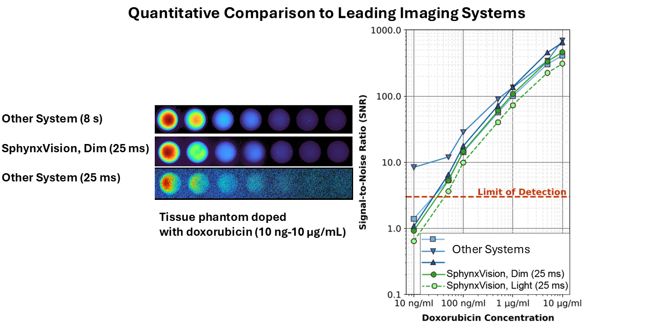
SphynxVision is designed to make preclinical fluorescence imaging easier to access, easier to repeat, and easier to integrate into active experimental workflows.
Move fluorescence imaging closer to the researcher instead of moving every study through a shared closed-chamber system.
Support interventions, dosing, heating, surgical access, perfusion, and device-assisted studies without interrupting the workflow.
Capture live fluorescence response and export ROI, intensity, and kinetic data for analysis.
SphynxVision is designed for studies where imaging needs to happen during the experiment — not only after the animal has been moved into a closed imaging chamber.
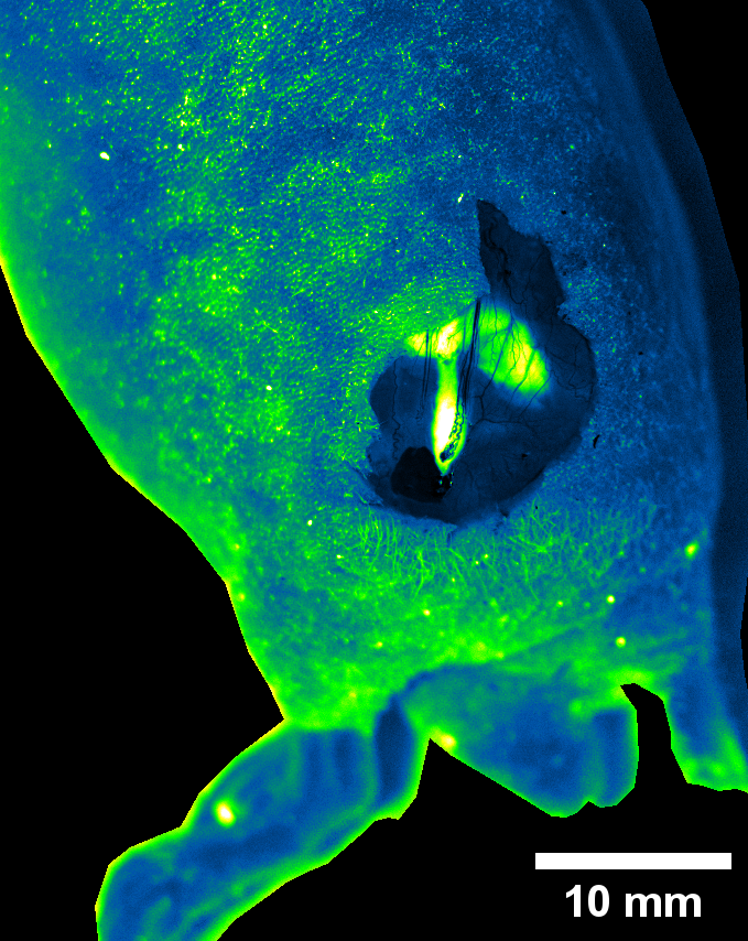
Real-time fluorescence visualization during tumor localization, surgical manipulation, or tissue-level intervention.
Closed chamber systems limit physical access and make it difficult to image while the procedure is actively changing.
SphynxVision keeps the imaging field open, allowing researchers to intervene and visualize fluorescence response at the same time.
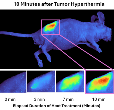
Monitor fluorescence changes during thermal activation, triggered release, nanoparticle delivery, or thermosensitive liposome studies.
Drug release and uptake can happen quickly. Endpoint imaging may miss when and where delivery occurred.
SphynxVision enables fluorescence imaging during heating or triggering, helping researchers capture release kinetics in real time.
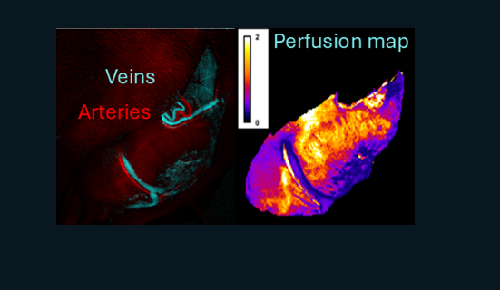
Track vascular dynamics, perfusion maps, angiography, and kinetic fluorescence changes across time.
Perfusion and vascular response can change rapidly and may not be captured by a single endpoint image.
SphynxVision supports dynamic imaging and downstream ROI or kinetic analysis for vascular and perfusion-focused studies.
Haemmerich, D., Newton, D.A., Patel, R.P., Rossmann, C. & VanDenburgh, A. Dynamic Contrast-Enhanced Fluorescence Imaging of Tumors with an Open Imaging System. World Molecular Imaging Congress (WMIC), Salt Lake City, UT, USA (2026).
Haemmerich, D., Newton, D.A., Patel, R.P., Rossmann, C. & VanDenburgh, A. An Open Preclinical Imaging System Enables Fluorescence Image-Guided Procedures. World Molecular Imaging Congress (WMIC), Salt Lake City, UT, USA (2026).
Haemmerich, D., Rossmann, C., Newton, D.A., Patel, R.P. & VanDenburgh, A. Fluorescence imaging system for intra-procedural preclinical applications. IEEE Engineering in Medicine and Biology Conference (EMBC), Toronto, Canada (2026).
Haemmerich, D., VanDenburgh, A., Rossmann, C., Newton, D.A. & Patel, R.P. Imaging chemotherapy delivery from thermosensitive liposomes by fluorescence imaging. Society for Thermal Medicine Annual Meeting, Savannah, GA, USA (2026).
Haemmerich, D., VanDenburgh, A., Rossmann, C., Newton, D.A. & Patel, R.P. Fluorescence imaging system for real-time visualization of chemotherapy delivery. World Molecular Imaging Congress (WMIC), Anchorage, AK, USA (2025).
SphynxVision brings together imaging science, biomedical engineering, and translational research experience to help researchers visualize dynamic fluorescence workflows in real time.

CEO & Founder
Dieter Haemmerich serves as CEO and Founder and is faculty at the Med. Univ. of South Carolina. He served in various leadership roles in two prior biomedical start-up companies, one of which was acquired by Johnson & Johnson in 2016. He co-authored more than 100 papers and book chapters covering the areas of biomedical imaging, image-guided therapies, computer modeling and instrumentation. He holds 8 patents, two of which have been exclusively licensed by Medtronic, with a commercial device based on his technology commercially available and clinically used since 2004 to treat more than 10,000 cancer patients. He brings deep imaging and therapy expertise to the SphynxVision platform.
View LinkedIn profile
CTO
Christian serves as CTO and is a multi-time founder and technology leader with a PhD in computational science and a background in advanced simulation. His experience includes finite element modeling, patient-specific thermal therapy simulations, additive manufacturing simulation, and technology commercialization. For SphynxVision, he contributes computational, software, and systems-level product strategy.
View LinkedIn profile
VP, Technical Operations
Krishna Ramajayam serves as VP of Technical Operations at OncoBLAZE and brings experience spanning academic research, biotechnology product development, and translational biomedical engineering. Prior to OncoBLAZE, he worked at Unchained Labs, where he contributed to the development and commercialization of six biotechnology products in less than three years, spanning lipid nanoparticles, extracellular vesicles, viral vectors, and biopharmaceutical characterization. His research encompasses image-guided drug delivery, liposomal therapeutics, hyperthermia, biomedical imaging, and computational modeling. He has authored or co-authored more than 25 journal publications and conference proceedings. He brings extensive translational research and industry product-development experience to the SphynxVision™ platform.
View LinkedIn profile


